


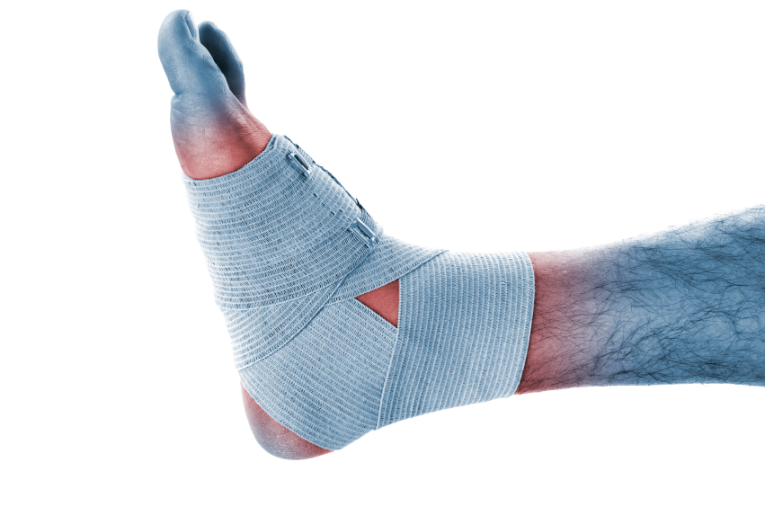
Anyone who has worked with field and court sport athletes has undoubtedly dealt with his fair share of athletes with ankle injuries. The ankle is the most frequently injured joint in sport, accounting for one-third of all injuries. As the Western approach to medicine is highly reactionary in nature, we typically follow ankle injuries up with rest and taping to assist the body in stabilizing motion. Unfortunately, in many cases, this isn’t enough to restore proper function at the ankle and leads to a loss of ankle dorsiflexion and an increased likelihood of repeated ankle injury in the future.
Why more dorsiflexion?
Sports are extremely stressful to the body, and the ankle joint is no exception. Over the course of an athletic practice or competition, each foot contacts the ground hundreds or thousands of times and goes into plantar flexion for propulsion equally as many times. This repetitive strain can shorten the plantar flexors, and over time, jarring of the joint causes natural gliding to become restricted. Add to this mix the fact that the ankle is the most commonly injured joint in sport along with countless taping jobs for ankle instability and elevated heel shoes and you get a nasty cocktail of muscular and joint restrictions that limit dorsiflexion.
A loss of dorsiflexion creates an inability to actively dissipate force in the lower extremity and can lead to problems both locally and up the chain, as the force must go somewhere. Furthermore, poor ankle dorsiflexion can lead to a “bouncy” appearance of the gait, as the body’s weight is transferred to the forefoot prematurely leading to inefficient locomotion.
Ankle anatomy and mechanics
Like most things, to get an understanding of how to fix an issue, you must first understand the proper mechanism. In reality, when talking about the ankle joint, we’re really speaking about two different joints—the “true ankle joint” consisting of the tibia, fibula, and talus and the subtalar joint including the talus superiorly and calcaneus inferiorly.
Traditionally, the true ankle joint is responsible for dorsiflexion and plantar flexion movements while the subtalar joint makes pronation and supination possible. As is often the case when discussing anatomy, it isn’t nearly as simplistic to state that dorsiflexion only occurs at the true ankle joint. Instead, there is a complex interplay of other, smaller joints in the foot that are to some extent controlled by motion at the “big boy” subtalar and true ankle joints.
The midtarsal joint—consisting of calcaneocuboid and talonavicular joints—also plays an important role in dorsiflexion and understanding how it works is essential to understanding a very common compensation pattern with restricted dorsiflexion. The midtarsal joint consists of two axes of motion—the oblique and the longitudinal axis. Of utmost consideration to this article is the oblique axis of motion because it allows a large amount of movement to occur including dorsiflexion and abduction. The oblique axis of the midtarsal joint has a one to one ratio of abduction and dorsiflexion. This means that for every one degree the joint abducts, one extra degree of dorsiflexion is created.
Herein lies the problem. This additional dorsiflexion created at the midtarsal joint is only possible with increased pronation at the subtalar joint. Thus, you’ll often observe overpronation and abducted feet in those lacking good dorsiflexion range of motion at the true ankle joint. This compensation is extremely common because we are very much a pronation dominated society on account of our footwear choices and the subsequent weakening of the feet. This poses a threat to the athletes’ well-being as a number of injuries have been associated with overpronation including plantar fasciitis, Achilles’ tendinopathy, patellofemoral pain, metatarsal stress fractures, and more. Clearly then, getting dorsiflexion from the oblique axis isn’t a good way to gain dorsiflexion if longevity is a concern.
So how should it be achieved? At the true ankle joint first and foremost. At this joint, dorsiflexion occurs by having the talus glide posteriorly into the ankle mortise. In cases of repeated ankle sprains, this posterior glide becomes increasingly small. It has been hypothesized that this occurs because of the talus’ lack of muscular attachments, which makes anterior subluxation easier following damage to its ligamentous attachments.
The ankle higher up
While the traditional approach sees an ankle injury as a problem specifically disturbing function at the foot and ankle, this isn’t necessarily the case. Those with repeated incidence of ankle sprain and functional ankle instability present a unique series of circumstances. It’s quite common for those with functionally instable ankles to exhibit low back pain.
In an attempt to figure out exactly why, Marshall and colleagues tested time to stabilization and trunk muscle activity in those with functional ankle instability (FAI) and a control group. The researchers found that the FAI group had delayed time to stabilization and delayed activation of the trunk musculature. Interestingly, there was no difference between the control group and the FAI group’s vertical jump heights. Is it a good thing that we are jumping our athletes and letting them participate in full sport activity despite an inability to reduce force properly?
Interestingly, Todd Wright of the University of Texas has said repeatedly, “If the ankle and foot are tight, there is a good chance that the core ain’t right.” I tend to agree. The foot and ankle are extremely important in driving the body, and any dysfunction at that level may lead to poor mechanics up the chain. It may manifest as low back pain, knee pain, or even shoulder pain, but regardless of its location, it could very well be the ankle that is causing the issue. One may also wonder if we’re getting optimal results from our athletes’ “core” training without first restoring the function of the ankle.
Fixing the issues
As the issues can be both joint mobility restrictions and muscular tightness, achieving proper dorsiflexion should be addressed with a multifaceted approach including altering tissue lengths, joint mobility, and other modifiable lifestyle factors.
Tissue length
While tissue length can be obtained with general static stretching, I’ve begun to favor an eccentric heel drop protocol for improving this quality. This decision is for several reasons.
1. Series elastic compliance: In stretching a muscle that is actively contracting, you aren’t only able to stretch the parallel elastic component but also the series elastic component. This in effect can allow greater gains in range of motion and tissue length changes.
2. Connective tissue strength: It has been demonstrated that eccentric heel drop exercises can improve tensile strength of the Achilles tendon, which is simply a bonus of this type of protocol that I feel presents some preventative benefit as well.
3. Increased sarcomeres in series: Eccentric exercise has been associated with increases in sarcomeres in series. It is theorized that this increase allows the muscle to be protected from stress, as each individual sarcomere would have to lengthen less in a series of 10 versus a series of five sarcomeres to achieve the same overall muscle length change.
4. FAI benefits: A loss of dorsiflexion accompanies repetitive ankle injuries, and athletes exhibiting FAI demonstrate reduced eccentric plantar flexion strength. To me, anything that can help change this is important, and anything that can improve strength to expedite a return to full function is a great thing to have in the program.
It’s important to note that in a heel drop series, the exercise should be performed both with an extended knee and with a flexed knee to preferentially engage both gastrocnemius and soleus, respectively.
Another exercise I really enjoy is a loaded backward walk.
Joint mobility
The traditional wall ankle mobilization is effective but can only go so far. Occasionally, you’ll find that it can even be contraindicated in some populations, as it may aggravate problems associated with anterior impingement of the ankle. For optimum results, it is wise to go back to the mechanics of the joint in learning how to mobilize it properly. If you recall, the talus is supposed to glide posteriorly relative to the tibia and fibula with dorsiflexion, so by pulling the lower leg anteriorly, we help facilitate the appropriate actions.
In this video, you can see that the athlete has fixed the heel and drives the knee over the toe exactly as a traditional wall ankle mobilization. It’s important to keep the athlete from driving into pronation. It’s also important to keep the knee in line with the second toe to make sure we’re getting mobility in the right places, not reinforcing poor movement quality.
Lifestyle factors
It’s imperative to also reduce the negative influences in everyday life in restoring joint function.
The foot is designed to provide a stable base upon which to push off and to accept the loads of the body during movement. Unfortunately, the modern shoe creates a significant hurdle in developing and maintaining this stability.
Typically, the modern shoe has an elevated heel, which shortens the tissues on the posterior side of the lower leg, making the restoration of proper length even more difficult. By providing a safe housing for the foot, modern shoes prevent the intrinsic foot muscles that are neurological triggers for muscle activation up the chain from firing at a high level. Combine this with thousands of foot contacts per day, and we’ve got a huge problem.
In an effort to prevent this, I try to get my athletes out of their shoes as much as possible. We very frequently do our warm ups unshod to allow the proprioceptor-dense feet to interface with the ground and restore some of the natural function. In some cases, I’ll attempt to progress to some running unshod or in shoes that offer great flexibility and little to no cushion, but this is done on a case by case basis and is implemented in a judicious manner.
Putting it all together
The foot and ankle are an extremely complex system that makes effective locomotion possible. In its complexity, however, I hope you can see that there are practical strategies that can help optimize its performance in sport and life. I hope that you’ve taken away from this article a better understanding of the ankle and its function as well as a practical approach to improving its motion.
Resources
- Behnke Robert S (2006). Kinetic anatomy. Champaign, Ill: Human Kinetics.
- Brukner Peter, Khan Karim (2006). Clinical Sports Medicine (McGraw-Hill Sports Medicine). Boston: McGraw-Hill Book Company.
- Butterfield Timothy A (2005). “Differential Serial sarcomere number adaptations in knee extensor muscles of rats is contraction type dependent.” Journal of Applied Physiology 1352–58.
- Duclay Julien, Martin Alain, Duclay Alice (2009) “Behavior of fassicles and the myotendinous junction of human medial gastrocnemius following eccentric strength training.” Muscle & Nerve 39:819–27.
- Fox Docherty, Applegate, Schrader (2008) “Eccentric plantar-flexor torque deficits in participants with functional ankle instability.” Journal of Athletic Training 43:51–54. Print.
- Mahieu, Nele, McNair, Cools (2007) “Effect of Eccentric Training On the Plantar Flexor Muscle-Tendon Tissue Properties.” Medicine and Science in Sports and Exercise 117–23.
- Marshall, McKee, Murphy (2009). “Impaired Trunk and Ankle Stability in Subjects with Functional Ankle Instability.” Medicine and Science in Sports and Exercise 1549–57.
- Siff Mel Cunningham (2003). Supertraining. Annapolis: Supertraining Institute.
- Vicenzino Bill, Branjerdporn Michell (2006) “Initial Changes in Posterior Talar Glide and Dorsiflexion of the Ankle after Mobilization with Movement in Individuals with Recurrent Ankle Sprain.” Journal of Orthopedic and Sports Physical Therapy 36(7):464–71.