The continuing saga of; aging, injuries, training setbacks and life.
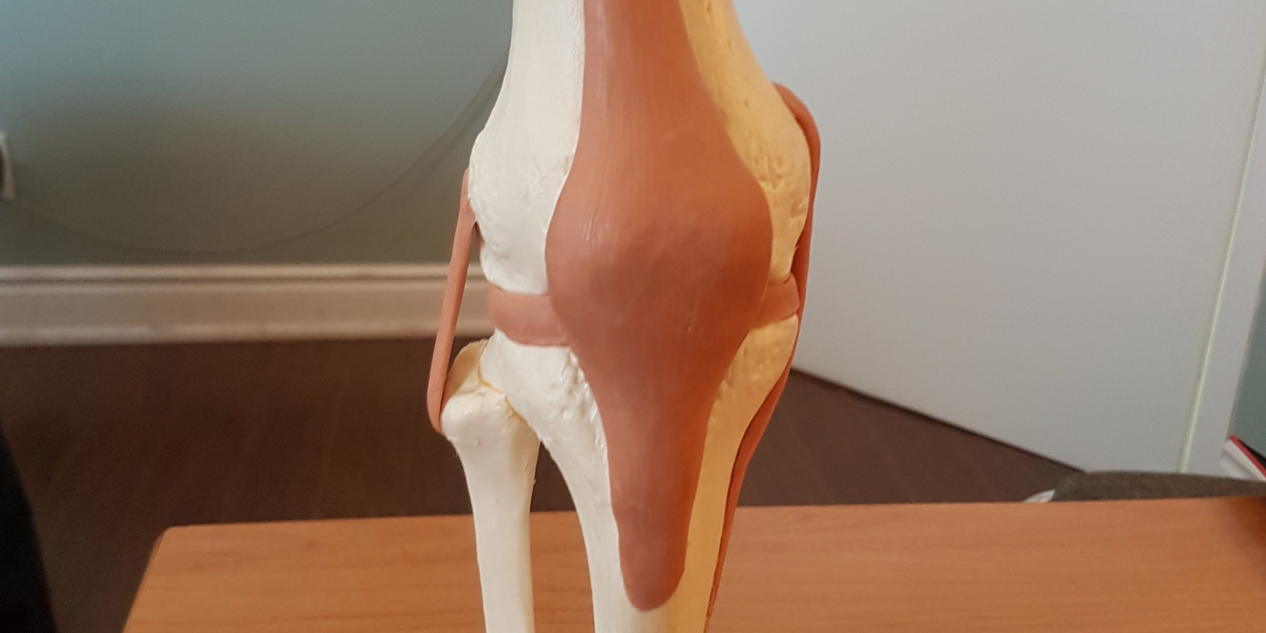
I have known for years my knee is getting worn out. The Dr. always said knees either wear out or rust out. Three or four weeks ago it flared up so painful I could not walk. I went to the Dr. for some tests to ensure I hadn't torn anything and that I was still able to train for the 21 Deadlifts Salute. Here are my results.
X-RAY RESULTS
Stage 3 OA is classified as “moderate” OA. In this stage, the cartilage between bones shows obvious damage, and the space between the bones begins to narrow. People with stage 3 OA of the knee are likely to experience frequent pain when walking, running, bending, or kneeling. They also may experience joint stiffness after sitting for long periods of time or when waking up in the morning. Joint swelling may be present after extended periods of motion, as well.
ULTRASOUND RESULTS
At this time, the patellar and quadriceps tendons remain intact. A moderate amount of fluid is seen in the suprapatellar bursa associated with some synovial thickening but no vascularity. The medial collateral ligament is slightly thicker than on the left side and there is also some tenderness in this area with transducer pressure suggesting a mild tendinosis. The lateral collateral ligament of the right knee appears normal. There is no evidence of a Baker’s cyst. The popliteal vessels appear satisfactory.
CONCLUSION:
Moderate quantity of fluid in the surapatellar bursa associated with some synovial thickening but no increased vascularity and there is an appearance indicating mild tendinosis of the medial collateral ligament.
In view of the nature of the injury and the patient’s past history, I would recommend an MRI assessment of the right knee if pain does not resolve fairly quickly to exclude meniscal injury.
MRI RESULTS (Finally saw the Dr. today)
I don't want to bore you with the long drawn out medical garble so here is the bottom line.
There is moderate degeneration of the lateral femortibial compartment of the knee joint with loss of articular cartilage over the weight bearing surface of the lateral tibial plateau and the lateral femoral coddle. There is marked osteophytic lipping.
Osteophytic lipping of the posterior aspect of the tibial plateau is noted. A small trace of joint effusion is noted.
IMPRESSION:
Destruction of the anterior horn and the middle third of the lateral meniscus with degeneration of the lateral femorotibial compartment of the knee joint.
My Doctor kinda laughed when she read me the word destruction. She said she has never seen that word used in an MRI report before. Funny but not funny at the same time.
So now I'm being referred to an orthopedic surgeon. I guess then I will get real answers on moving forward and surgery options.
Until then, I am cleared to lift. My Dr. said and I quote "Go ahead and lift you won't damage it anymore than it already is".
I will continue to train carefully for the 21 Deadlift Salute but that will be my last so called "Kick at the Can" for a while. I will be taking a break after the Arnold's to have my knee fixed.
#injuriesuck #setbacksuck #driven #teamelitefts #painsucks






1 Comment